

Every scale of life is beautiful, big and small. Sometimes the splendor of life is hidden behind literal scales.
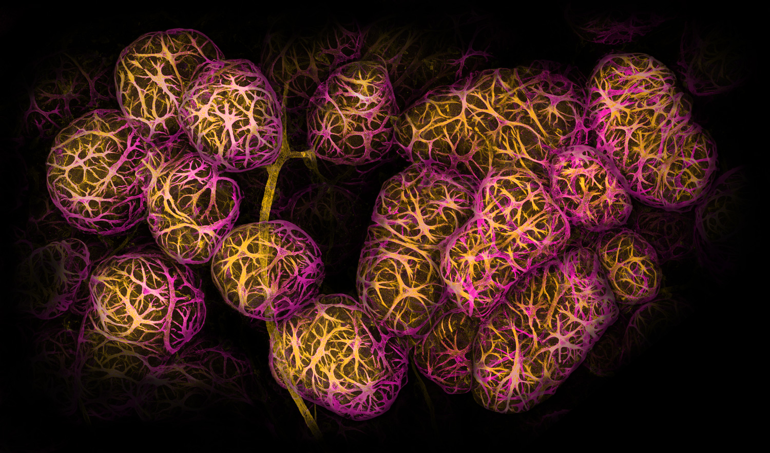
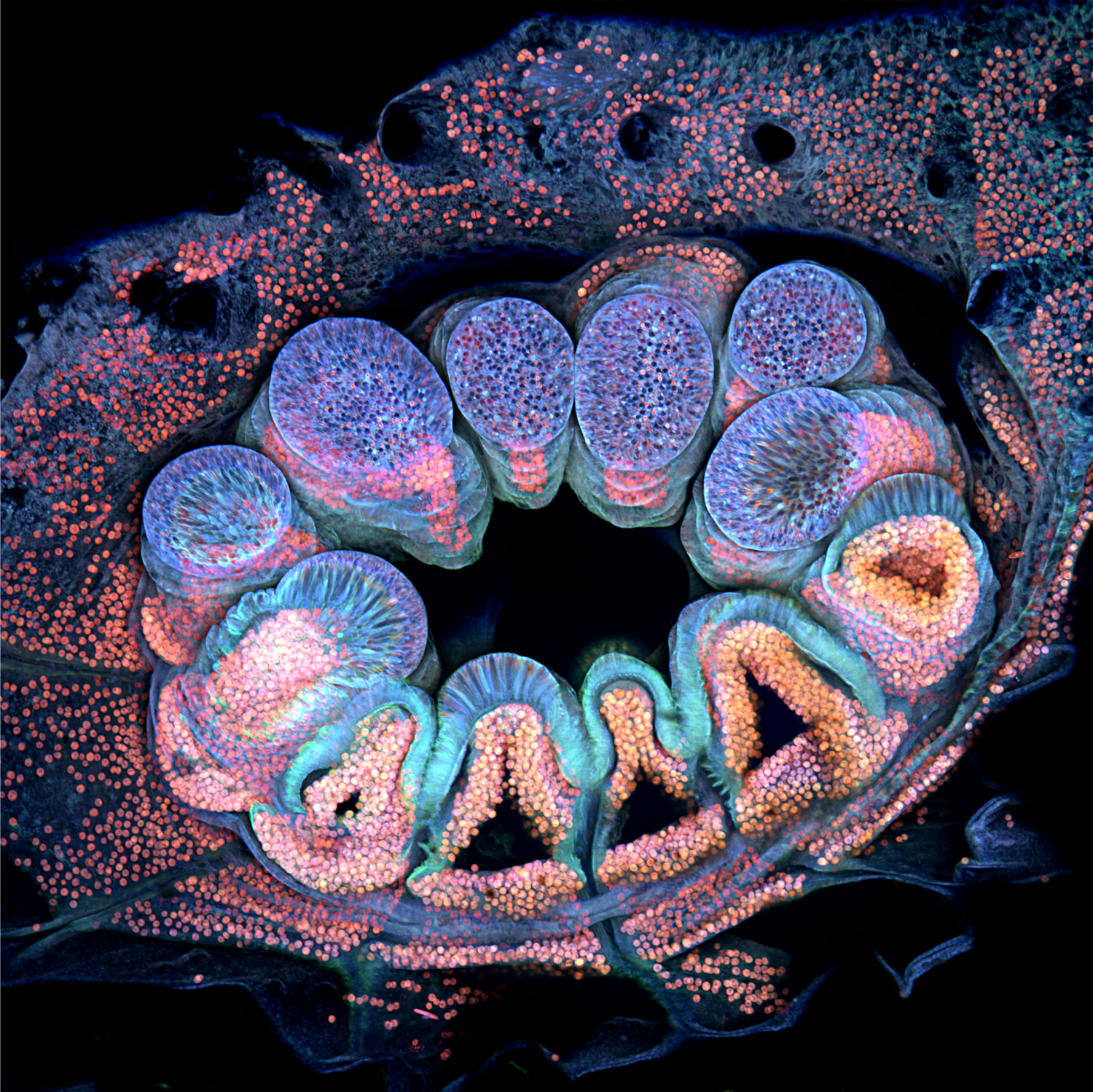
An amazing glimpse below the embryonic scales of a Madagascar giant daygecko (Phelsuma grandis(Won first place in The 2022 Nikon Small World Photomicrography Competition. Grigorii Timin of University of Geneva and Michel Milinkovitch crafted the winning image from hundreds of images captured over two days using a Confocal Microscope. They study the genetics as well as the physics of embryonic growth.
To highlight budding nerves in cyan, and structures containing collagen in a range o oranges and yellows, the hand is artificially dyed. Milinkovitch says collagen is the building block of all life. Research can be improved by understanding the exact location of collagen, which will help scientists better understand the development of tissues and bodies.
Timin says that parts of the bones that have begun calcifying shine brightest in this image. As orange branches, developing tendons and ligaments stretch. Blood cells form clusters or line up inside new blood vessels at the tips of the gecko’s digits.
Milinkovitch states that the image shows beauty in all sizes. The snapshot is “beautiful as a hand, you see this beautiful pattern of the fingers. You can zoom in to see the spongy bones. The tendons are visible when you zoom in. And you zoom, and you see the fibers that’s from the tendons. Then you zoom, and you see the blood cells.”
The gecko photo is one of 92 incredible images recognized in this year’s competition. The 48th annual contest’s winners AnnouncedOctober 12. These are just a few of our favorites.
Got milk?
The photograph looks like a cluster or grapes from a distance. Each orb is actually a clumping of cells within breast tissue.
Caleb Dawson of Parkville’s Walter and Eliza Hall Institute of Medical Research took thousands of images with a confocal microscopy to examine tiny, musclelike cells wrapped around milk-producing spheres. This image, which won second place, was marked with dyes and antibodies.
Dawson says the hormone oxytocin stimulates the cells. The hormone oxytocin is released when breastfeeds and helps extract milk from the spheres called alveoli. Researchers can use such images of breast tissue to help them understand how immune cells protect breast tissue and the babies that it can feed.
Snuffed candle
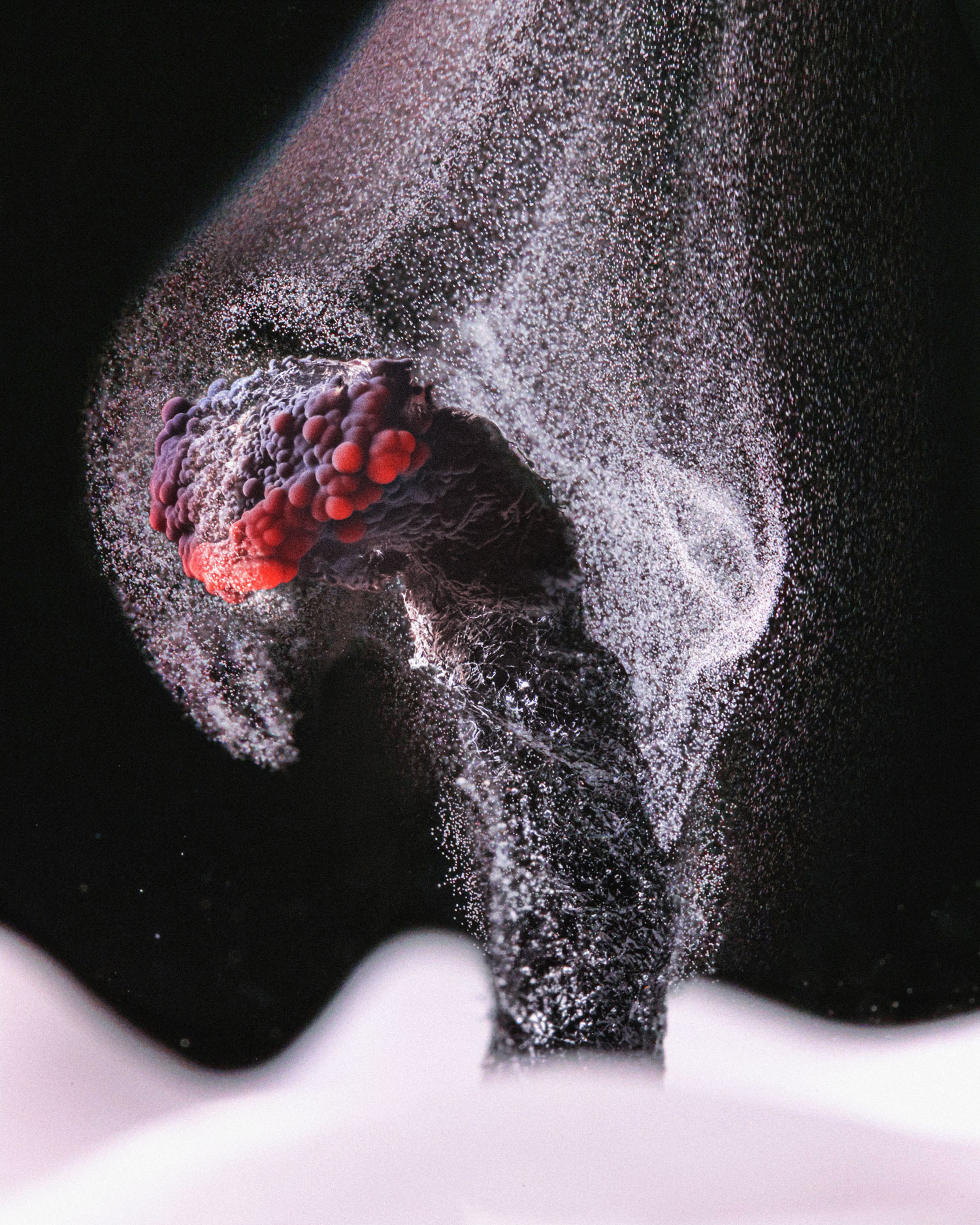
Ole Bielfeldt needed to grab the last candle flame quickly.
The main components of candle wax are hydrogen and carbon atoms. When they’re lit, most turn into carbon dioxide. Some hydrocarbons do not burn. Instead, they collect as soot on surfaces around the candle. “When the flame goes out, the glowing wick has enough heat left to break up the wax molecules for a while, but not enough to burn the carbon,” says Bielfeldt, a photographer in Cologne, Germany. “So you get a trail of smoke until it cools.”
Bielfeldt captured the unburned carbon particles drifting away using a fast shutter speed with a bright LED lamp. He earned sixth place.
Iridescent slime mold
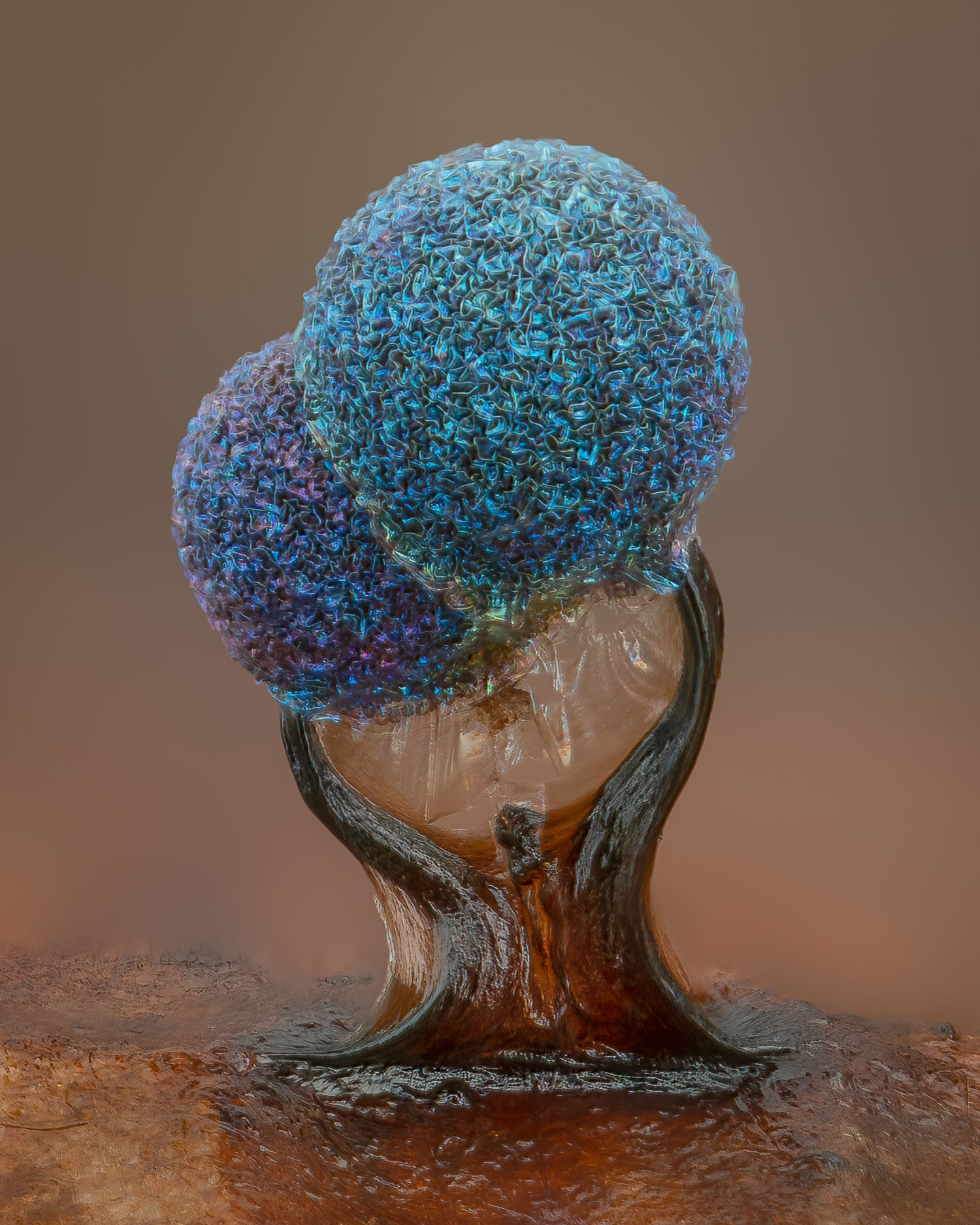
These are tiny pieces of art that can be hidden on leaves or decaying logs in moist forest. Lamproderma Slime molds
Alison Pollack from San Anselmo, Calif. saw a sparkling leaf while she was working in a leaf pile under the warm October sun. After taking the leaf home, Pollack was captivated by the crinkly heads of slime mold and the iridescence. Pollack was able to capture a striking photo after about 40 hours and 147 combined shots later. This photo won fifth place in the contest.
Slime molds are known for their smooth heads that release spores into their environment. Pollack believes that the rapid drying of this pair might have slowed their development and left their heads wrinkled. But that’s OK, “because the texture to me is just gorgeous.”
A deadly predator
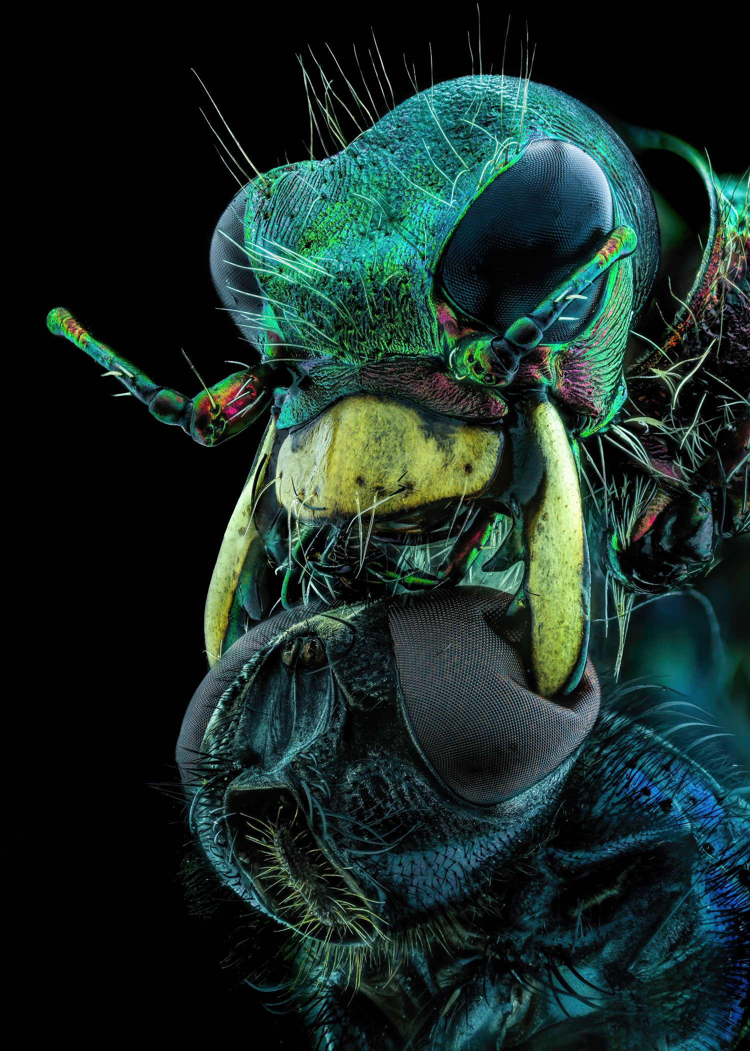
Everyone fears the predacious Tiger Beetle, particularly this poor fly.
Murat Öztürk of Ankara, Turkey, nabbed 10th place in this year’s competition with an astounding — and unnerving — snapshot of a tiger beetle using its mandibles to crush a fly by its eyes.
Tiger beetlesCicindelinae() Sprint after their prey so fast that they are Insects go temporarily blind. The beetle in the photo would have stopped several times to orient its body to find the fly, before eventually taking its dinner. Thanks to the beetle’s strong and sharp jaws, “the chances of survival of the creatures caught by this insect are very low,” Öztürk says.
Coral close-up
Some cauliflower coral can be found off the coast Australia’s Opal Reef.Pocillopora verrucosaPolyps can appear green. The same organism can be transformed under a microscope in a laboratory.
To reveal the polyp’s individual cells, marine scientist Brett Lewis of Queensland University of Technology in Brisbane, Australia, stitched together more than 60 images taken over 36 hours. When exposed to different wavelengths light, coral fluoresces a variety of colors including pinks, purples, and blues. Algae living inside the polyp appear orange or pink, while the coral’s tissues shine blue. The competition awarded the image 12th place.
Lewis said that the photo shows algal cells shining through a blue haze in certain areas. That’s because coral tissue is transparent; algae give coral its color.
Peeks of coral’s internal makings can help scientists understand its biology, Lewis says. His work, for instance, aims to figure out how young polyps build strong foundations when they attach to a surface — an important step in building or restoring coral reefs.
