

‘Portray’ with fluorescent markers has lengthy been a useful solution to detect distinctive double-stranded constructions in DNA. As soon as restricted to a palette of simply 256 colours, scientists can now create gorgeous works of laboratory artwork with unimaginable 16 million shades and hues.
The brand new approach precisely recreates digital pictures with 24-bit colour depth, and the outcomes are breathtaking.
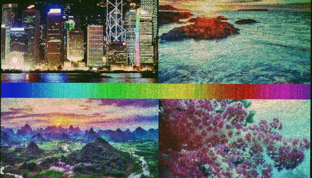
Here is an authentic digital picture:
And the DNA ‘painted’ model:
It is not only a new inventive format. The miniaturized DNA-based portray strategy is a scale-up on microarray know-how for learning gene expression, and it has different potential too.
Researchers usually use artworks to check or showcase strategies which will later show helpful in real-world functions.
“Past imaging, a DNA colour code may have very helpful functions in information storage on DNA,” says chemist Tadija Kekić from the College of Vienna.
Changing information into DNA sequences saved on a chip is much like storing data in a barcode. This new methodology by Kekić and one other College of Vienna chemist, Jory Lietard, may enable a better quantity of storage on a smaller floor.
Their approach is so exact it might be used to color micrometer-scale options onto biopolymers. Doable makes use of embody biosensors and diagnostics, the place fine-tuned management of DNA self-assembly is essential.
An enormous quantity of knowledge may be saved in DNA as a code consisting of sequences made up of 4 chemical bases – adenine, guanine, cytosine, and thymine. Every base additionally corresponds to a associate, seeing to the formation of complementary sequences within the type of a double strand.
Having one sequence in hand offers scientists a software for locating its matching associate in a jumbled mess. Analytical strategies based mostly on DNA sequences caught to a strong floor in a grid sample, known as DNA microarrays, depend on coloured fluorescent markers to point out when complementary DNA strands bind collectively.
This course of the place complementary DNA strands acknowledge one another and bind collectively as double strands is called hybridization. Residing organisms use the straightforward, secure guidelines of hybridization to learn and duplicate their genetic data.
To visualise how we will flip fluorescent DNA sequences into vibrant artworks, it’d assist to image the way in which trendy colour shows, like telephones and laptop computer displays, create a variety of colours.
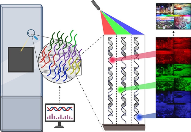
The colour of every pixel within the grid of your display is made up of crimson, inexperienced and blue main channels, and the depth of every channel may be dialed up or down to supply the colour you see.
Having mismatches between bases on a double strand primarily provides a small quantity of instability to its hybridization, altering the molecular construction of double-stranded DNA.
The researchers behind this new fluorescent hybridization approach programmed instability into the strands to alter the brightness of their fluorescent markers. By utilizing completely different dyes for various colours and eradicating bases, hybridization on patterned DNA surfaces could make patterns which might be very easy to see.
Particular dyes (Cy3, Cy5, and fluorescein) on fragments of DNA, known as probes, create 256 shades of sunshine in every of the crimson, inexperienced, and blue channels. This creates a slider-like fluorescence sign between not one of the colour and full colour.
“Basically, our synthesis floor turns into a canvas for portray with DNA molecules on the micrometer scale,” says Lietard.
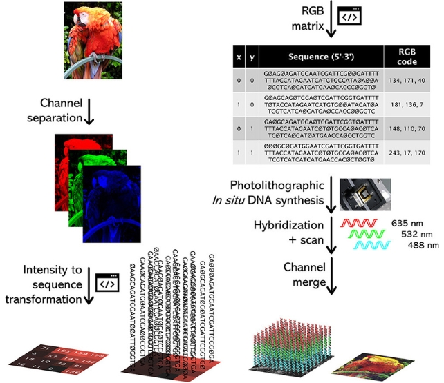
To exhibit the colour vary, the group separated digital pictures into three 8-bit RGB layers and assigned DNA sequences for every pixel’s worth. On a fingernail-sized microarray, the approach – maskless array synthesis mixed with photolithography – can synthesize lots of of 1000’s of distinctive DNA sequences directly.
With the assistance of a machine with lots of of 1000’s of tiny mirrors similar to the pixels within the picture, and pc scripts, greater than 786,000 DNA sequences can match on the microarray floor in a grid formation. That is one for every pixel unit of 14 x 14 micrometers within the RGB layer, with data on the depth worth encoded within the DNA phase.
Scanning the three tiny microarrays, or exactly organized ‘DNA canvases’, after which merging the layers again collectively digitally, permits copy of the unique picture in additional than 16 million colours with a decision of 1024 x 768 pixels. The group thinks the method might be expanded to work at full HD and even 4K.
“Digital pictures may be reproduced with excessive constancy on the micrometer scale by utilizing a easy course of,” the authors write.
Amongst many makes use of, larger decision fluorescence indicators may result in extra exact measurements of processes occurring inside our our bodies, permitting higher understanding of cell biology, and earlier detection of ailments like most cancers.
The research has been printed within the Journal of the American Chemical Society.
