

Scientists believe that bone marrow cells within the skull respond differently to diseases, which could make the skull a good way to track and treat inflammation of the brain.
Researchers from Germany and UK who studied this reaction have proposed that their findings may be used as a noninvasive skull imaging method.
This could be a game changer for neurological diseases. You can find out more about this by clicking here. neuroscientist Ali Ertürk from Munich University in Germany.
This breakthrough could allow for more accurate monitoring of conditions like Alzheimer’s“Early detection can prevent strokes and other diseases, including heart attacks.”
Neuroinflammation The brain and nervous system are affected by many diseases.It activates immune cells, and releases inflammatory molecules to heal and protect our tissues. But it comes at a price. risking damageCompromise healing. The skull and other membranes cover the brain making it difficult to reach the area for treatment.
Scientists are working to find out how they can help. Recently discoveredThe protective membranes on the outermost surface of brain, which is the bone marrow, form pathways. meningesThe skull and brain can interact directly, allowing immune cells to move.
It’s widely known that Immune cells can enter brainAfter strokes and other problems, the Blood-brain barrierIt is unclear how immune cells access the brain layers through the skull, and how frequently they do so compared to the other routes.
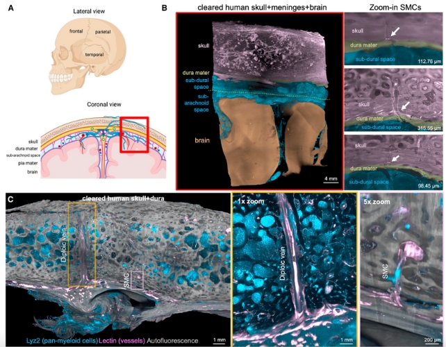
The team combined 3-D imaging with tissue clearing to visualize samples of human brain, skull, and meninges.
Tissue ClearingThe process of making biological tissues transparent. This process allows light to pass through brain tissue and skull for microscopic examination.
Researchers observed that the skull-meninges connection (SMCs), which extends closer to the surface of the brain than previously thought, penetrates the dura mater, the outermost and most tough meninges membrane.
These findings are profound and suggest that the connection between the brain and skull is more complex than previously believed. You can find out more about this by clicking here.Ilgin Kolabas from the University of Munich is a Neuroimmunologist.
frameborder=”0″ allow=”accelerometer; autoplay; clipboard-write; encrypted-media; gyroscope; picture-in-picture; web-share” allowfullscreen>
Kolabas and his colleagues also studied six different bones to study cells. Dura materDifferent bones, including the brain, have different molecular profile, with immune cells unique to the skull.
A protein analysis of postmortem human skull, spine and pelvic bones samples revealed the unique molecular profiles. The CalvariaThe genes and cellular receptive proteins that were most differentially expressing in this area, mostly related with migration and inflammation, were located here.
The team found that both mouse and human skulls had specialized cells. neutrophilCells, a white blood cell type that is crucial to the body’s defense against infection.
Using a type of functional imaging known as positron emission tomographyThe team discovered that (PET) detected changes in skull signals that mirror the signals from the underlying human brains of Alzheimer’s patients and stroke victims. The team also detected an increase of disease-specific translocator proteinIn many neurological diseases, (TPSO), signals are present in various parts the skull.
frameborder=”0″ allow=”accelerometer; autoplay; clipboard-write; encrypted-media; gyroscope; picture-in-picture; web-share” allowfullscreen>
Scientists believe that their discoveries regarding the immune response of the skull could be used to detect brain inflammation using a simple scan of a person’s skull.
“This could potentially be done using portable and wearable devices, offering a more accessible and practical way to monitor brain health,” Ertürk Explaining.
Alzheimer’s disease and stroke are examples of neurological diseases that affect millions around the globe.
The team said that “our detailed demonstration of inflammation of the skull in various diseases in humans could be used to diagnose or monitor diseases in the future.” Close“But detailed clinical trials are required to explore its clinical usefulness.”
The journal has published the study Cellular.
