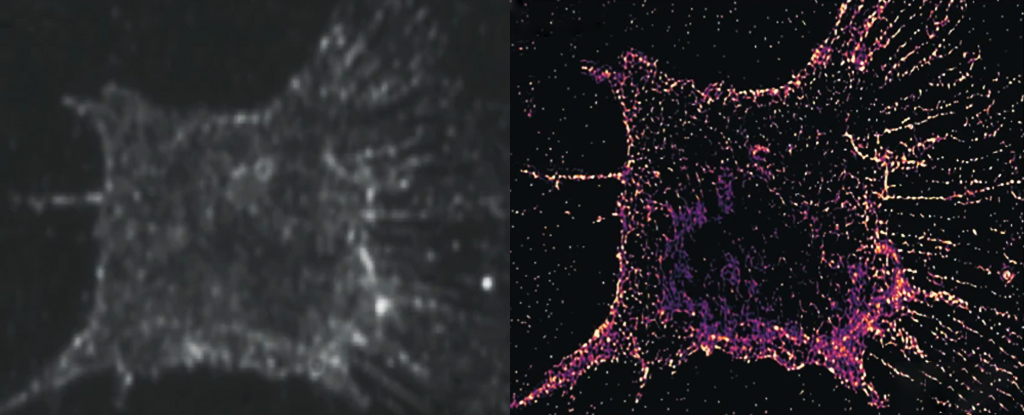
Researchers have discovered a new level of detail within living cells by replacing fluorescent molecules with those that scatter light.
The new tweak allows scientists to monitor molecular behavior for a longer time period. It opens a window into crucial biological processes, such as cell division.
The living cell is an active place, with proteins bouncing around. explainsGuangjie Cui, biomedical engineer at the University of Michigan. “Our superresolution allows us to see these dynamic activities in a very appealing way.”
It is possible to observe extremely small biological structures using superresolution. It takes a series snapshots of fluorescing molecule constellations that highlight selected areas of the tissue. This eliminates the blurring effects of a flood diffracted by light.
Researchers who developed it won a Nobel prize in 2014. As revolutionary as this process was, it is the ability of the molecules fluorescing to absorb and then spit the Required wavelength of LightThe mapping of processes lasting longer than a few seconds is impossible because the sensor wears out in a matter of seconds.
Cui and his colleagues instead developed a method to detect the light scattered off of randomly distributed gold nanorods, a technique that does not break down after repeated exposure. Although the gold markers were larger than target structures, imaging different subsets from the rods in a variety of angles and combining images provided the same level of detail.
The system allows for 250 hours of continuous observations with a resolution as low as 100 atoms.
Cui and his colleagues examined the entire division process with their PINE nanoscopy. They discovered a previously unknown behavior of actin moleculesThe molecule-level analysis is a key component of the.
The major component of the cell’s structure is actin. cytoskeletonThis molecule provides structural support to cells and facilitates movement within the cell. These branching filament-shaped molecules are crucial in dividing cells and separating them into two daughter cell.
Due to limitations in our visual technology, it has been difficult to determine how these cells are identical.
Cui and his group observed 904 filaments of actin during the process of cell division to determine how molecules interacted with one another. When actin molecules were less bound together, they expanded in search of new links. As each actin reaches out to its neighbors, other actin molecules are drawn closer, increasing the network.
Researchers were able to see how these smaller scale movements translated into a larger cellular scale. Unexpectedly the cell actually contracts when actin is expanded, but expands when this occurs. Researchers are interested in understanding how this opposite motion occurs.
Somin Lee, a biomedical engineer at the University of Michigan, said: “We will use our method to examine how other molecular components organize into organs and tissues.” WriterThe Conversation
Our technique may help researchers better understand the molecular defects that can develop in organs and tissues.
The research has been published by Nature Communications.


